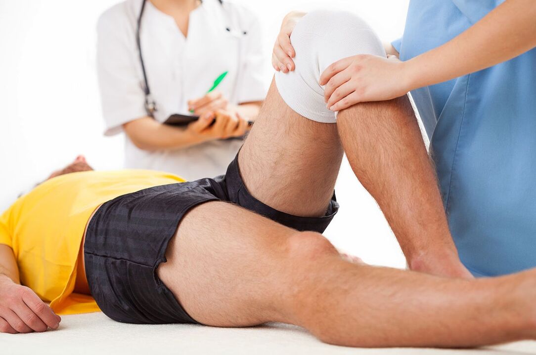
Arthropathy is the most common joint disease. According to experts, 6. 43% of the population in my country suffers from this disease. Both men and women often suffer from arthropathy, however, males have a slight advantage in younger patients and females in older patients. An exception to the general situation is arthropathy of the interphalangeal joint, which is 10 times more common in women than in men.
With age, the incidence rate increases sharply. Therefore, according to research, arthropathy has been detected in 2% of people under 45 years of age, 30% of people 45 to 64 years old, and 65-85% of people 65 years of age and older. Arthropathy of the knee joint, hip joint, shoulder joint and ankle joint has the greatest clinical significance due to its negative impact on the patient's living standard and working ability.
reason
In some cases, the occurrence of this disease has no obvious cause, and this joint disease is called idiopathic or primary.
There is also secondary arthropathy-which develops due to certain pathological processes. The most common causes of secondary joint disease are:
- Injury (fracture, meniscus injury, ligament rupture, dislocation, etc. ).
- Dysplasia (congenital dysplasia of joints).
- Degenerative malnutrition process (Perthes disease, osteochondrotis dissecans).
- Diseases and conditions that involve increased joint mobility and weakness of ligament organs.
- Hemophilia (arthropathy due to frequent blood in the joints).
Risk factors for developing joint disease include:
- Getting older.
- overweight
- Excessive pressure on the joints or specific joints.
- Surgical intervention on joints,
- Genetic predisposition (arthropathy in close relatives).
- Endocrine disorders in postmenopausal women.
- Neurodystrophy diseases of the cervical or lumbar spine (shoulder arthritis, lumbar iliac syndrome).
- Repetitive minimal trauma to the joints.
Onset
Arthropathy is a multi-cause disease, regardless of the specific cause, it is based on the destruction of the normal formation and recovery of cartilage tissue cells.
Generally, articular cartilage is smooth and elastic. This allows the articular surfaces to move freely relative to each other, providing the necessary shock absorption, thereby reducing the load on adjacent structures (bones, ligaments, muscles, and joint capsules). For arthropathy, the cartilage becomes rough, and the articular surfaces begin to "adhere" to each other during exercise. The cartilage is getting looser. The small pieces are separated from it, fall into the joint cavity and move freely in the joint fluid, damaging the synovium. On the surface area of the cartilage, small calcification foci appear. In the deep layers, ossification areas appear. Cysts are formed in the central area and communicate with the joint cavity. Around the joint cavity, due to the pressure of the intra-articular fluid, an ossification area is also formed.
Pain syndrome
Pain is the most persistent symptom of arthropathy. The most significant signs of joint pain are the connection with physical activity and weather, night pain, initial pain, and sudden severe pain combined with joint block. With prolonged exercise (walking, running, standing), the pain will increase, and the pain will subside during rest. The cause of night pain in arthropathy is venous congestion and increased blood pressure in the bones. Unfavorable weather factors can exacerbate the pain: high humidity, low temperature and high air pressure.
The most typical symptom of arthropathy is initial pain-the pain that occurs during the first exercise after a break and disappears when the exercise is maintained.
symptom
Arthropathy gradually develops. Initially, patients worry that mild, short-term pain that is not clearly located will be aggravated by physical exertion. In some cases, the first symptom is a crunching noise when moving. Many patients with arthropathy experience joint discomfort and short-term stiffness during the first exercise after a period of rest. Later, night and weather pain supplemented the clinical manifestations. As time goes by, the pain becomes more and more obvious, and the movement is obviously restricted. As the load increased, the joint on the opposite side began to be injured.
The exacerbation period and the remission period alternate. The exacerbation of joint disease usually occurs in the context of increased stress. Due to pain, muscle reflex spasm of the limbs can form muscle contractures. The contraction of the joints becomes more and more constant. Muscle cramps and muscle and joint discomfort occur during rest. Limp occurs due to increased joint deformity and severe pain syndrome. In the later stages of arthropathy, the deformity becomes more obvious, the joints are bent, and the movement in them is obviously restricted or non-existent. Support is difficult; arthropathy patients must use a cane or crutches when moving.
diagnosis
The diagnosis is made on the basis of typical clinical symptoms and X-ray images of arthropathy. Take X-rays of the diseased joints (usually two projection methods are used): X-rays of the knee joint, X-rays of the knee joint, hip joint disease-X-rays of the hip joint, etc. X-rays of arthropathy consist of physical signs of dystrophic changes in articular cartilage and adjacent bone areas. The joint space is narrowed, the bones are deformed and flattened, cysts are formed, subchondral bone sclerosis and osteophytes appear. In some cases, arthropathy will show signs of joint instability: bent limbs, subluxation.
Taking into account the radiological signs, experts in the field of orthopedics and traumatology distinguish the following stages of arthropathy (Kellgren-Lawrence classification):
- The first stage (suspected joint disease)-the joint space is suspected to be narrow, missing or fewer osteophytes.
- Stage 2 (Mild Arthropathy)-Suspected joint space narrowing, clearly define osteophytes.
- Stage 3 (moderate arthropathy)-The joint space is significantly narrowed, there are obvious osteophytes, and skeletal deformities may occur.
- Stage 4 (severe joint disease)-significant narrowing of the joint space, osteophytes, obvious bone deformities and bone sclerosis.
Sometimes X-rays are not enough to accurately assess the condition of the joints. In order to study bone structure, joint CT is performed to assess the state of soft tissue-MRI of the joint.
treat
The main goal of treating patients with arthropathy is to prevent further cartilage destruction and maintain joint function.
During remission, patients with arthropathy receive physical therapy. The set of exercises depends on the stage of joint disease.
Medications for the worsening stage of joint disease include the use of non-steroidal anti-inflammatory drugs, sometimes in combination with sedatives and muscle relaxants.
Long-term use of arthropathy includes chondroprotective agents and synovial prostheses.
In order to relieve pain, reduce inflammation, improve microcirculation and eliminate muscle spasms, patients with arthropathy are referred for physical therapy. In the exacerbation stage, laser therapy, magnetic field and ultraviolet radiation are prescribed, and in the remission stage-electrophoresis with dimethylamine, tramecaine or novocaine, ultrasound with hydrocortisone, induction heating, thermal procedures (groundWax, paraffin), sulfide, radon and sea bathing. Perform electrical stimulation to strengthen muscles.
If the articular surface is damaged and there is obvious joint dysfunction, arthroplasty is performed.


